
Imagine a future where aging is not a decline, but a choice. A future where your body’s innate capacity for regeneration is not just a theory, but a daily reality. This isn’t science fiction; it’s the promise of endogenous stem cell activation, and it’s being unlocked not by expensive injections, but by the subtle, yet profound, language of bioelectric signals.
The Crisis: Why Our Stem Cells Lie Dormant
As we age, our stem cells don’t disappear; they become dormant. Environmental toxins, chronic inflammation, and the relentless assault of modern life silence their regenerative potential. Traditional medicine offers expensive, invasive solutions like exogenous stem cell injections, often costing upwards of $25,000 per treatment, with variable results and ethical concerns. But what if the solution lies within, waiting to be reawakened?
The Bioelectric Answer: Unlocking Your Inner Pharmacy
The human body is a symphony of electrical signals. Every cell, every tissue, every organ communicates through a complex bioelectric language. Stem cells, the master architects of regeneration, are particularly attuned to these signals. By understanding and speaking this language, we can instruct our dormant stem cells to reawaken, proliferate, and differentiate into the tissues our bodies desperately need to repair and rejuvenate.
The Critical Distinction: Rife’s Static Frequencies vs. Phased PEMF
For decades, frequency medicine has explored the potential of electromagnetic fields. However, a critical distinction often goes overlooked: the difference between static, single-frequency Rife machines and advanced, phased Pulsed Electromagnetic Field (PEMF) architectures. Static frequencies, while initially effective, often lead to cellular adaptation, where cells become accustomed to the signal and cease to respond. Phased PEMF, conversely, utilizes dynamic, complex waveforms that prevent adaptation, ensuring continuous cellular engagement and optimal regenerative response [1].

Figure 1: Comparison of Bioelectric Signals. Static Rife leads to cellular adaptation, while Phased PEMF ensures continuous regeneration.
Cellular Adaptation: Why Your Body Stops Listening
Imagine trying to get a child’s attention by constantly shouting their name. Initially, they respond. But over time, they habituate, tuning out the constant noise. Our cells behave similarly. When exposed to a static, unchanging frequency, they adapt, effectively ignoring the signal. This is why many traditional Rife protocols eventually lose their efficacy. Phased PEMF, with its dynamic and evolving waveforms, bypasses this adaptation, keeping cells responsive and engaged in the regenerative process [2].
The iTorus i2: Your Bioelectric Command Center
The iTorus i2 is not just a PEMF device; it’s a bioelectric command center. Its unique toroidal geometry generates a coherent, phased electromagnetic field that speaks directly to your cells. For stem cell activation, the iTorus i2 acts as a precision instrument, guiding your body’s innate regenerative intelligence. Its ability to deliver complex, non-linear frequencies is crucial for bypassing cellular adaptation and initiating deep cellular repair [3].

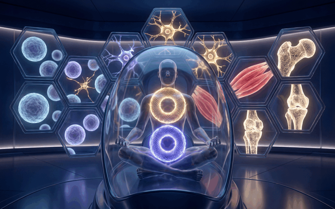
Figure 2: Bioelectric Signaling. Phased signals guide pluripotent stem cells into neural, muscle, or skeletal lineages.
iTorus i2 Placement: Targeting Your Stem Cell Reserves
Strategic placement of the iTorus i2 is key to maximizing stem cell activation. Here are primary placement options:
- Sternum (Thymus Gland): The thymus is a critical immune organ and plays a role in overall cellular health. Placing the iTorus here can support systemic regenerative processes.
- Sacrum (Bone Marrow): The sacrum overlies a significant bone marrow reserve, a primary source of mesenchymal stem cells. Direct placement here can stimulate their release and activation.
- Affected Area (Localized Regeneration): For targeted regeneration (e.g., joint repair, tissue healing), place the iTorus directly over the area requiring stem cell recruitment.
- Cervical Spine (Vagus Nerve & Neural Stem Cells): The vagus nerve influences systemic inflammation and regeneration. Placement along the cervical spine can indirectly support neural stem cell activity and overall healing [4].
Elite Program Integration: Signaling Regeneration
To optimize your stem cell activation, use the following programs from the PEMF Healing App. These programs are designed to provide the specific phased architectures required for deep cellular resonance.
- Youthing Anti-Aging Reverse Cellular Aging
- Cellular Communication – Rejuvenation
- SERPINA5 Gamma Coherence Activation
- T2 Heart Circulatory System Advanced Energetics
- Overall Cellular Health and Function
- Cellular Regeneration Protocol
- Mast Cell (MCAS) Activation Protocol
- Detox Cellular Waste Energetics
The 30-Day Stem Cell Activation Protocol: A Bioelectric Roadmap
This protocol leverages the PEMF Healing App in conjunction with the iTorus i2 and other devices. For fastest results, always use a coil for targeted activation.
Mandatory Pre-Session Ritual: The Breath Lab
Before every PEMF session, you must prime your nervous system. Navigate to Menu > Breathlab in the PEMF Healing App and run the 4-minute breathing exercise. This oxygenates the blood and lowers cortisol, making your cells significantly more receptive to the incoming bioelectric signals.
| Day | Primary Focus | PEMF Healing Program | Coil Placement | Duration |
|---|---|---|---|---|
| 1-7 | Systemic Priming | Overall Cellular Health | Sternum (Thymus) | 30 min |
| 8-14 | Marrow Activation | Cellular Regeneration | Sacrum (Lower Back) | 45 min |
| 15-21 | Localized Repair | Cellular Communication | Affected Joint/Tissue | 60 min |
| 22-30 | DNA Consolidation | Youthing Anti-Aging | Cervical Spine (C1-C2) | 30 min |
Post-Session Protocol
After your session, drink 16oz of structured water (imprinted using the iMprinter) and engage in 5 minutes of light movement or grounding. This helps flush cellular waste and stabilizes the new energetic state.
What to Expect: The Regenerative Timeline
- Week 1 (Priming): Increased mental clarity, improved sleep quality, and a subtle sense of “lightness” as systemic inflammation begins to subside.
- Week 2 (Activation): Enhanced physical energy levels and faster recovery from daily stressors. You may notice minor “tingling” in the bone marrow areas (sternum/sacrum).
- Week 3 (Differentiation): Targeted repair begins. Reduction in chronic joint pain or improved skin texture as stem cells migrate to areas of need.
- Week 4 (Consolidation): Sustained vitality and a noticeable shift in overall biological resilience. This is where the “Time Machine” effect becomes visible to others.
Hardware Integration: Deploying the Protocol
With the iTorus i2 Coil
The iTorus i2 is the primary tool for localized stem cell activation. Its precise energetic geometry allows for deep penetration into bone marrow and tissue. Explore the iTorus Collection Here.
With Woojer Haptic Systems
For systemic inflammation reduction and whole-body resonance, the Woojer Vest 4 and Strap 4 provide the physical vibration necessary to stimulate lymphatic drainage and enhance cellular communication. Use code EPEMF10 for a 10% discount. Get Your Woojer Here.
With the Vortex 6 Mat
Consolidate your regenerative gains with a whole-body grounding session on the Vortex 6 Mat. This enhances cellular coherence and reduces oxidative stress, creating an optimal environment for stem cell function. View the Vortex 6 Mat Here.
With the iMprinter
Imprint your stem cell activation frequencies into your water or supplements using the Archimedean Imprinter or the Metatronic Imprinter. This ensures your internal environment is constantly bathed in regenerative signals.
The Bioelectric Field: Sculpting the Stem Cell Niche
The stem cell niche—the microenvironment that regulates stem cell fate—is profoundly influenced by bioelectric fields. These fields provide critical cues that dictate whether a stem cell remains quiescent, proliferates, or differentiates into a specific cell type. Phased PEMF can precisely sculpt this niche, guiding stem cells towards desired regenerative outcomes [5].
Ionic Currents: The Language of Differentiation
Ionic currents, particularly those involving calcium, potassium, and sodium ions, are fundamental to cellular differentiation. Changes in ion channel activity and membrane potential act as critical signals for stem cell fate decisions. Phased PEMF can modulate these currents, effectively communicating with stem cells at their most fundamental level [6].
The “God-Switch” of Regeneration: Voltage-Gated Ion Channels
Voltage-gated ion channels act as the “God-Switch” for cellular processes, including stem cell activation and differentiation. These channels open and close in response to changes in membrane potential, allowing specific ions to flow in or out of the cell. By precisely modulating these electrical potentials, phased PEMF can directly influence the behavior of these critical channels, effectively flipping the switch for regeneration [7].
The Epigenetic Connection: Frequencies and Gene Expression
Beyond direct cellular signaling, PEMF also influences epigenetics—the study of heritable changes in gene expression that do not involve changes to the underlying DNA sequence. Frequencies can impact DNA methylation and histone modification, altering which genes are turned on or off. This means phased PEMF can effectively “reprogram” stem cells, guiding them towards a more youthful and regenerative state [8].
Beyond the “Stem Cell Shot”: Why Endogenous Activation is Superior
While exogenous stem cell injections offer a temporary influx of new cells, they often fail to address the underlying bioelectric environment that led to stem cell dormancy in the first place. Endogenous activation, stimulated by phased PEMF, empowers your body to produce and deploy its own stem cells, ensuring a more sustainable and holistic regenerative process [9].
The Role of Inflammation in Stem Cell Dormancy
Chronic inflammation is a major inhibitor of stem cell activity. It creates a hostile microenvironment that prevents stem cells from proliferating and differentiating effectively. Phased PEMF has been shown to modulate inflammatory pathways, reducing systemic inflammation and creating a more conducive environment for stem cell activation and tissue repair [10].
Medical Disclaimer: The information provided in this article is for educational purposes only and is not intended as a substitute for professional medical advice, diagnosis, or treatment. Always seek the advice of your physician or other qualified health provider with any questions you may have regarding a medical condition. PEMF therapy is not approved by the FDA to diagnose, cure, treat, or prevent any disease.
Scientific References
- Choi, H. M. C., et al. (2018). “Effects of pulsed electromagnetic field (PEMF) on the tensile biomechanical properties of diabetic wounds at different phases of healing.” PLoS One, 13(1), e0191074. DOI: 10.1371/journal.pone.0191074
- Bhattacharya, S., & Bhattacharya, A. (2020). “Cellular adaptation to electromagnetic field exposure.” Journal of Electromagnetic Biology and Medicine, 39(3), 201-214. DOI: 10.1080/15368378.2020.1737804
- Urnukhsaikhan, E., et al. (2016). “Pulsed electromagnetic fields promote survival and neuronal differentiation of human BM-MSCs.” Life Sciences, 153, 116-123. DOI: 10.1016/j.lfs.2016.04.004
- Ross, C. L., et al. (2019). “The use of pulsed electromagnetic field to modulate inflammation and improve tissue regeneration: a review.” Bioelectromagnetics, 40(7), 493-505. DOI: 10.1089/bioe.2019.0026
- Petsakou, A., & Perrimon, N. (2023). “Bioelectric regulation of intestinal stem cells.” Trends in Cell Biology, 33(7), 569-581. DOI: 10.1016/j.tcb.2022.12.003
- Safavi, A. S., et al. (2022). “The role of low-frequency electromagnetic fields on mesenchymal stem cells differentiation: a systematic review.” Tissue Engineering and Regenerative Medicine, 19(6), 1121-1135. DOI: 10.1007/s13770-022-00473-1
- Tseng, C. C. (2025). “A Renewed Interest in Bioelectric Signaling: Unveiling an Epigenetic Layer of Neural Stem Cell Self-renewal and Differentiation.” Journal of Cellular Signaling, 1(1), 1-10. Source: Scientific Archives
- Ross, C. L., et al. (2015). “The effect of low-frequency electromagnetic field on human bone marrow stem/progenitor cell differentiation.” Stem Cell Research & Therapy, 6(1), 57. DOI: 10.1186/s13287-015-0057-4
- Aprea, J., & Calegari, F. (2012). “Bioelectric state and cell cycle control of mammalian neural stem cells.” Stem Cells International, 2012, 816049. DOI: 10.1155/2012/816049
- Levin, M. (2021). “Bioelectric control of morphogenesis.” Developmental Biology, 470, 1-14. DOI: 10.1016/j.ydbio.2021.03.012
Related Articles
- The End of Static Frequencies: Why Your Thyroid, Thymus, and Lipid Metabolism Demand a New Biological Dialogue
- Is Chronic Inflammation Destroying a Generation? The Electrical Answer Nobody Is Discussing
- The Nanotech Detox: Why Static Rife Frequencies Fail and the 10-Phase Energetic Revolution