
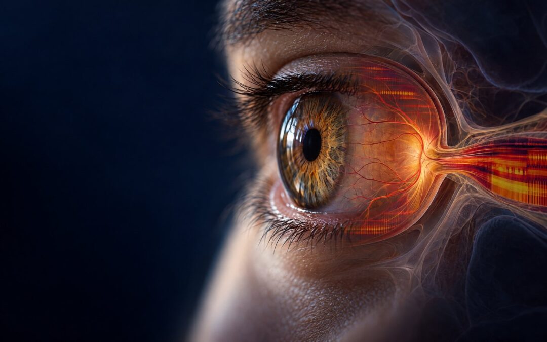
Glaucoma is not one signal. It is a clinically monitored optic-nerve disease with pressure, anatomy, retinal ganglion-cell biology, blood-flow questions, and visual-field change all in the picture. That complexity is exactly why a responsible ocular research guide has to begin with the science readers can act on today: regular eye care, prescribed treatment, and clear separation between established glaucoma management and experimental signal-design ideas.
Glaucoma & Optic Nerve BioPhi Matrix 12-Phase BioPhi-Harmonic Ocular Research Energetics is presented here as a structured ocular-research and listening framework. It organizes distinct research topics into an evolving 12-phase sound-and-signal experience. It is not an established glaucoma treatment, an eye-pressure intervention, an optic-nerve regeneration protocol, or a replacement for an ophthalmologist’s monitoring plan.
The practical value of this guide: understand the biology and research language behind the program, inspect the supplied spectrogram accurately, keep clinical care in the lead, and use a consistent, non-clinical routine without turning a consumer program into a claim about intraocular pressure, vision, or disease progression.
The exact program and its place in a responsible ocular research workflow
The featured public program is Glaucoma & Optic Nerve BioPhi Matrix 12-Phase BioPhi-Harmonic Ocular Research Energetics. Its value as a program guide is in its organization: it keeps multiple ocular-research themes inside one clearly named, time-structured experience rather than collapsing glaucoma into a single phrase or a single implied mechanism.
That organization does not establish a clinical effect. Studies of ocular electrical stimulation and historical ocular electromagnetic exposures used particular medical hardware, tissue contact or field delivery, timing, and clinical measurements. Those conditions cannot be assumed to transfer to headphones, haptics, consumer coils, an app, or an audio-led routine.[6][7][8] The right conclusion is more useful than a bigger claim: the program can be discussed as a carefully organized research experience while glaucoma care remains clinician-led.
| Program layer | What is linked | Responsible reader use |
|---|---|---|
| Featured guide | Glaucoma & Optic Nerve BioPhi Matrix 12-Phase BioPhi-Harmonic Ocular Research Energetics | Use as a research-oriented listening and signal-design guide. Do not use it to self-treat glaucoma or alter prescribed care. |
| Related public catalogue | Glaucoma Energetics; Optic Nerve Diseases Energetics; Eye Health – Recovery | Explore exact public pages as catalogue references. Program titles are not clinical evidence. |
| Additional eye-topic pages | Eye Strain Energetics; Retinal & Ocular System, Isochronic 7-Phase Vision Restoration Energetics; Vision Restore: Retina & Macula 9-Phase Bilateral Toroidal Energetics | Keep one program at a time when taking non-clinical comfort notes. Do not combine titles into a claim about visual restoration. |
| Further catalogue context | Eyesight – Vision Disorders Energetics; Eye Floater Basic Energetics; Dry Eye – Syndrome – Recover Energetics | Different eye complaints and diseases need different clinical evaluation. A broad catalogue is not a substitute for diagnosis. |
Why the optic nerve requires a multi-domain research lens
Glaucoma is a group of eye diseases that can damage the optic nerve. It may progress slowly, often without early symptoms, which is why comprehensive dilated examinations and visual-field testing matter.[1] The American Academy of Ophthalmology describes clinical monitoring as a combination of pressure assessment, optic-nerve and retinal-nerve-fiber-layer evaluation, structural imaging, and functional visual-field testing.[2]
Retinal ganglion cells are the output neurons of the retina. Their axons travel together to form the optic nerve, carrying visual information onward toward the brain. Research investigates mechanical stress, vascular factors, oxidative stress, inflammatory signaling, impaired axonal transport, and other pathways that may shape retinal ganglion-cell vulnerability.[3] A 12-phase research architecture can therefore be useful as an editorial map of these distinct questions. It should never be mistaken for 12 verified clinical interventions.

The established anchor: pressure lowering and scheduled monitoring
For primary open-angle glaucoma, lowering intraocular pressure is the established treatment target. The AAO guideline notes that treatment goals are individualized and that visual fields and optical coherence tomography provide complementary information for following change over time.[2] The National Eye Institute lists prescription eye drops, laser treatment, and surgery among standard treatment options.[1]
This creates a constructive way to use a research guide. Keep the things that are already proven to matter visible: attend appointments, use prescribed drops as directed, communicate side effects, and bring questions about any new wellness practice to the treating eye-care team. The research framework can help a reader stay engaged with the science, but it cannot supply a pressure reading, assess an optic nerve, or replace a visual-field test.
What the submitted spectrogram actually shows
The supplied image is a spectrogram, also called a time-frequency signal display. It visibly contains stacked horizontal bands, repeated changes in relative density, and brighter amber blocks that appear and recede across the timeline. Those are useful observations about the program’s evolving audio architecture. The image does not show an ocular dose, intraocular-pressure change, magnetic field at the eye, optic-nerve response, or treatment outcome.

The same distinction applies to the visible layering. A phased arrangement can make a long-form piece feel more varied and can give each chapter a recognizable design role. It does not by itself establish that the program prevents physiological habituation, changes retinal ganglion-cell biology, or outperforms a static signal. Those are separate questions that require direct, device-specific clinical research.

Why medication and substance themes appear in an ocular research atlas
The program’s research framing names substance and medication themes because they organize real areas of ocular science. They are not presented here as audio equivalents, molecular signatures, dosing instructions, or hidden treatment claims. Their purpose is to keep the reader oriented to the biology and pharmacology that researchers actually investigate.
Aqueous-humor and pressure-management context. Latanoprost and pilocarpine are examples of medication contexts associated with helping fluid drain from the eye. Dorzolamide, timolol, and brimonidine represent medication classes used to reduce the amount of fluid the eye makes. Acetazolamide and methazolamide are also clinical pressure-management medications in selected circumstances.[4] Naming these substances in a research atlas can clarify why aqueous dynamics and pressure remain central glaucoma topics. It does not mean a sound, haptic pattern, coil, or associated program can reproduce drug action.
Oxidative-stress and retinal-resilience context. Glutathione, N-acetylcysteine, vitamin C, curcumin, acetyl-L-carnitine, and GABA appear as research-language themes around redox biology, neuroinflammation, cellular energy, and retinal signaling. Oxidative stress and inflammatory pathways are active areas of glaucoma research, but evidence for nutritional supplementation in clinical glaucoma care remains mixed and controversial.[5] The responsible public discussion is therefore biological context, not a supplement protocol and not a frequency disclosure.
Confidentiality and accuracy: This article intentionally omits internal substance-associated values, derived sequences, carriers, modulation settings, and design calculations. A named substance is a research reference point, not evidence that a program reproduces its pharmacology.
What ocular electrical and electromagnetic research can contribute to the conversation
There is a genuine research literature on electrical and electromagnetic stimulation in ocular settings. It is compelling precisely because it is specific. A historical study of pulsed electromagnetic exposure in open-angle glaucoma used specialized ocular hardware and a defined multi-session protocol.[6] A transpalpebral electrical-stimulation pilot used current-controlled stimulation through the eyelid, while a transcorneal pilot used corneal electrodes and repeated clinical testing.[7][8]
These papers offer research context, not a transferable home routine. A small randomized transcorneal electrical-stimulation trial reported no statistically significant visual-field differences between groups and stated that people with primary open-angle glaucoma should receive that intervention only under study conditions.[9] A later retrospective analysis explored electrical neurostimulation in people already receiving intraocular-pressure-lowering treatment and explicitly identified the need for future randomized controlled trials.[10]
The useful takeaway is not that research should be ignored. It is that modality, hardware, tissue interface, dose, timing, outcome measurement, and study design matter. That specificity protects readers from turning a research title or a spectrogram into a clinical promise.
Three-day ocular research listening routine
This is a non-clinical routine for organized listening and general wellness journaling. It is deliberately limited to one exact program, so the reader can notice comfort and routine consistency without mixing multiple titles. It is not an eye-treatment plan. Do not place a coil on or near the eye or orbit. Do not use this routine to make changes to prescribed drops, laser plans, surgery plans, or clinical monitoring.
| Day | Program | What to record |
|---|---|---|
| 1 | Glaucoma & Optic Nerve BioPhi Matrix 12-Phase BioPhi-Harmonic Ocular Research Energetics | Use the platform’s standard supported playback method at a comfortable level. Note the date, setting, completion, and sound comfort only. |
| 2 | Glaucoma & Optic Nerve BioPhi Matrix 12-Phase BioPhi-Harmonic Ocular Research Energetics | Repeat in a similar quiet setting. Record the same non-clinical observations and any reason to stop for discomfort. |
| 3 | Glaucoma & Optic Nerve BioPhi Matrix 12-Phase BioPhi-Harmonic Ocular Research Energetics | Keep the experience consistent. Review only whether the routine felt acceptable and was completed as planned. Do not interpret comfort notes as eye-pressure, vision, or optic-nerve data. |
Device context and best practices
For this topic, the central best practice is conservative separation: a consumer wellness routine remains separate from eye care. The app can provide the linked program context. If a reader uses consumer hardware elsewhere in a general wellness practice, they should follow manufacturer instructions, use conservative settings, and keep it away from the eye and orbital area. There is no coil-placement protocol for glaucoma in this article.
| Layer | Optional resource | Appropriate role in this article |
|---|---|---|
| Program access | Frequency Healing App | Find the exact public program page and follow the platform’s supported workflow. |
| Consumer PEMF | iTorus i2 or iTorus i5 | General consumer-wellness context only. Do not position a coil at the eye or treat it as an ocular medical device. |
| Haptic layer | Woojer Vest 4 | Optional body-based listening layer, separate from ocular care and not a glaucoma intervention. |
| Daily-practice ritual | Metatronic Flower of Life Frequency Imprinter | Optional personal ritual tool only. Do not make health claims about imprinted water or use it in place of prescribed care. |
Simple daily practices support the clinical plan without competing with it: keep eye appointments, use prescribed drops exactly as directed, report side effects, carry an up-to-date medication and supplement list to visits, protect sleep and hydration, and ask an eye-care professional before adding a supplement or changing a wellness routine. The National Eye Institute specifically advises patients to discuss other medicines, supplements, and vitamins because glaucoma medicines can interact with them.[4]

When to seek urgent eye care
Sudden intense eye pain, a red eye, nausea, blurry vision, or halos around lights can be symptoms of angle-closure glaucoma and warrant urgent medical evaluation.[1] New or rapidly worsening vision change, a curtain-like shadow, severe headache with eye symptoms, or new neurologic symptoms also deserves prompt evaluation. Do not repeat a wellness session to test such symptoms.
Educational and wellness disclaimer: This article is for educational purposes only. It does not provide medical advice, diagnosis, or treatment, and it does not establish that any program or device will produce a particular ocular result. Do not delay or replace licensed medical care. Consult a qualified eye-care professional for symptoms, glaucoma, pregnancy, implanted electronic devices, medication questions, or any concern about whether PEMF is appropriate for you. Follow the manufacturer’s instructions for every device.
Related Articles
- The Evidence-Based Beginner’s Guide to PEMF Therapy
- Vascular Flow & Endothelial Support: Research and 8-Phase Guide
- The 20-Minute iTorus Muscle Test: 7 Days, Active vs Sham
References
- National Eye Institute. Glaucoma.
- American Academy of Ophthalmology. Primary Open-Angle Glaucoma Preferred Practice Pattern.
- Feng KM et al. The Role of Retinal Ganglion Cell Structure and Function in Glaucoma. Cells. 2023.
- National Eye Institute. Glaucoma Medicines.
- Pinazo-Durán MD et al. Strategies to Reduce Oxidative Stress in Glaucoma Patients. Curr Neuropharmacol. 2018.
- Tsiselskii IuV et al. The effect of a pulsed electromagnetic field on the hemodynamics of eyes with glaucoma. Oftalmol Zh. 1990.
- Gil-Carrasco F et al. Transpalpebral Electrical Stimulation as a Novel Therapeutic Approach to Decrease Intraocular Pressure for Open-Angle Glaucoma: A Pilot Study. J Ophthalmol. 2018.
- Ota Y et al. The Efficacy of Transcorneal Electrical Stimulation for the Treatment of Primary Open-angle Glaucoma: A Pilot Study. Keio J Med. 2018.
- Röck T et al. Transcorneal electrical stimulation in primary open angle glaucoma. Ophthalmologe. 2017.
- Erb C et al. Electrical neurostimulation in progressive glaucoma: a retrospective study. BMC Ophthalmol. 2022.