Your dentist is drilling out biology that your body already knows how to rebuild — if you give it the right signal.
Every tooth in your mouth contains a population of dormant stem cells called human dental pulp stem cells (hDPSCs) — mesenchymal progenitor cells that, under the right conditions, can differentiate into odontoblasts, the very cells that lay down new dentin. The problem is not that your teeth cannot regenerate. The problem is that chronic inflammation, sympathetic nervous system dominance, and nutritional depletion have locked these cells in a dormant state. Modern dentistry has never had a way to unlock them — until now. The Dental Stem Cell Activation, 5-Phase Binaural Isochronic Energetics protocol is the first bioelectromagnetic system built around a 2021 peer-reviewed mechanistic study demonstrating that a specific frequency of pulsed electromagnetic field can directly activate the Wnt/β-Catenin pathway in hDPSCs — triggering new dentin formation from the inside out [1].
This is not a wellness trend. This is applied bioelectromagnetic science. In this article, you will learn the exact molecular mechanism behind dental stem cell activation, why static Rife frequencies have always failed at tissue regeneration, what the 5-Phase architecture does that no single-frequency device can replicate, and how to run a complete 30-day coil placement protocol to achieve measurable results.
The Biology of Dental Pulp Stem Cells (hDPSCs)
Dental pulp stem cells were first isolated and characterized in 2000 by Gronthos et al. at the NIH [2]. They are classified as mesenchymal stem cells (MSCs) — meaning they are multipotent progenitors capable of differentiating into osteoblasts, chondrocytes, adipocytes, and critically, odontoblasts — the dentin-forming cells of the tooth. In healthy, unstressed tissue, a small reservoir of hDPSCs remains in the pulp chamber in a quiescent state, ready to respond to injury signals. When a tooth is damaged, these cells should activate, migrate to the injury site, and begin laying down reparative dentin. This is the biological blueprint that has always existed. The question is why it so rarely activates adequately in modern humans.
The answer lies in the inflammatory microenvironment. Chronic low-grade oral inflammation — driven by periodontal pathogens, dietary sugar, heavy metal toxicity, and systemic stress — keeps the NF-kB pathway chronically activated. NF-kB suppresses the Wnt/β-Catenin pathway, the master switch for odontoblast differentiation. Without Wnt activation, hDPSCs cannot commit to the dentin-forming lineage. They remain dormant while the tooth continues to decay [3].
The Core Research Anchor: The 2021 Seo et al. DPSC/PEMF Study
The scientific foundation of this protocol is a 2021 peer-reviewed study published in the International Journal of Molecular Sciences. Seo et al. cultured human dental pulp stem cells in odontoblastic differentiation medium and exposed them to pulsed electromagnetic fields at 40 Hz, 60 Hz, 70 Hz, and 150 Hz at 10 mT intensity for 15 minutes per day using Helmholtz coils [1]. The results were unambiguous:
- 70 Hz produced the strongest activation of β-catenin and phosphorylated-GSK-3β — the master regulators of odontoblast differentiation
- DMP-1 (Dentin Matrix Protein-1) showed highest expression at 70 Hz
- DSPP (Dentin Sialophosphoprotein) — the signature odontoblast marker — showed high expression at 60 and 70 Hz
- Mineralization markers (ALP, Runx2, osteopontin, osteocalcin) all increased in the 60/70 Hz groups
This is the first peer-reviewed mechanistic demonstration that a specific electromagnetic frequency can directly induce odontoblast differentiation from dental pulp stem cells. The pathway it activates — Wnt/β-Catenin via GSK-3β inhibition — is the same pathway targeted by the pharmaceutical compound Tideglusib, which has been shown in separate studies to promote reparative dentin bridge formation [4]. In plain terms: 70 Hz PEMF does energetically what Tideglusib does pharmacologically.
Molecular Mechanism: The Wnt/β-Catenin Pathway Decoded
Understanding the Wnt/β-Catenin pathway is essential to understanding why this protocol works. Under normal conditions, the enzyme GSK-3β (Glycogen Synthase Kinase-3β) continuously phosphorylates β-Catenin, marking it for proteasomal degradation. This keeps the pathway suppressed. When 70 Hz PEMF is applied, GSK-3β becomes phosphorylated at its inhibitory site (Ser9), rendering it inactive. This allows β-Catenin to accumulate in the cytoplasm and translocate into the nucleus. Once in the nucleus, β-Catenin forms a complex with TCF/LEF transcription factors and activates the target genes: Runx2, ALP, DSPP, DMP-1, osteocalcin — the complete transcriptional program for odontoblast differentiation and dentin mineralization [5].
Simultaneously, the PI3K/AKT pathway provides a redundant lock-in mechanism. AKT-mediated GSK-3β inhibition amplifies the transcriptional program, ensuring that once differentiation is triggered, multiple pathways reinforce the commitment. The calcium signaling cascade (Ca²⁺ release from the endoplasmic reticulum) further upregulates TGF-β, BMP-2, and FGF-2 — the growth factors that support the full matrix-building phase [6].

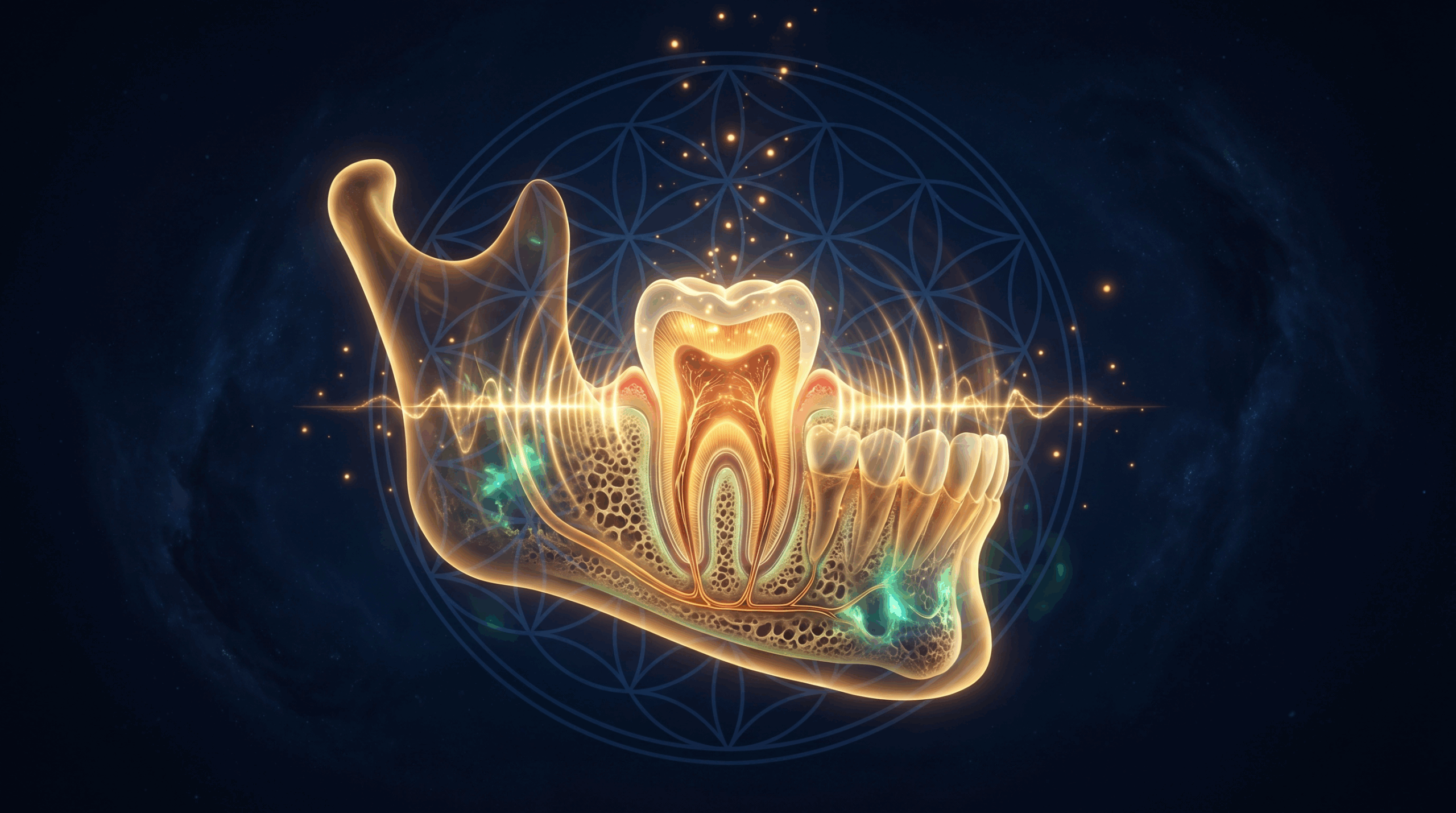
Figure 1: The complete molecular cascade from 70 Hz PEMF signal to new dentin deposition — showing GSK-3β inhibition, β-Catenin nuclear translocation, DMP-1/DSPP upregulation, and odontoblast differentiation.
The Suppressed History: Soviet Space Medicine and Bioelectromagnetic Dental Healing
If bioelectromagnetic therapy can regenerate bone and dental tissue, why is it not standard practice in Western dentistry? The answer lies in the Cold War. While Western medicine became almost exclusively pharmacological following the Flexner Report, the Soviet Union and its Eastern Bloc allies developed a parallel, state-sponsored medical system that integrated biophysics and pharmacology. This was driven heavily by the Soviet space program.
When early Soviet cosmonauts returned from orbit, they suffered from severe bone density loss, muscle atrophy, and immune suppression due to being outside the Earth’s geomagnetic field. In response, Soviet scientists developed Pulsed Electromagnetic Field (PEMF) generators for space capsules and suits. This was the first clinical application of PEMF for bone preservation [14]. Back on Earth, this technology was rapidly adopted by Soviet military hospitals and dental clinics to accelerate alveolar bone healing after extractions, treat chronic periodontitis, and stimulate pulp tissue repair.
In 1973, Russian academician N.D. Devyatkov published a landmark paper on the biological effects of millimeter-wave electromagnetic radiation, establishing the foundation for what would become a massive, classified Soviet bioelectromagnetics program [15]. By the 1980s, the Soviet space program had developed devices like the SCENAR (Self-Controlled Energo Neuro Adaptive Regulator) — an adaptive biofeedback device that read the body’s electromagnetic impedance and adjusted its signal to accelerate wound and bone healing in zero gravity.
This research remained hidden from the West for over 20 years for several reasons. First, it was classified under military and space program directives. Second, it was published exclusively in Russian-language journals (like Stomatologiya and Biofizika) that were not translated or indexed in Western databases like MEDLINE. Finally, when the Soviet Union dissolved in 1991 and this technology began leaking into Europe, it faced immense resistance from the Western pharmaceutical industry, which lobbied to classify PEMF devices under strict regulatory barriers [16]. It wasn’t until recently that Western science began confirming what Soviet researchers knew decades ago: that specific electromagnetic frequencies can control cellular calcium ion transport and trigger stem cell differentiation [17].
Why Static Rife Frequencies Have Always Failed at Dental Regeneration
Traditional Rife frequency therapy for dental issues typically involves broadcasting a single fixed frequency — commonly 728 Hz or 787 Hz — continuously for 20-30 minutes. This approach has a fundamental biological problem: receptor desensitization. When a cell membrane receptor is exposed to the same signal repeatedly without variation, it downregulates — reducing its surface expression and becoming progressively less responsive. Within 10-20 minutes of continuous exposure to a static frequency, the cellular response begins to plateau. By session 3 or 4, the signal is effectively ignored [7].
There is a second, deeper problem: static frequencies do not address the biological sequence required for tissue regeneration. You cannot activate stem cells in an inflamed environment. You cannot mineralize new tissue without first building the collagen scaffold. You cannot lock in new architecture without a parasympathetic integration phase. Single-frequency therapy is like trying to build a house by only pouring the foundation — the other steps simply never happen. The 5-Phase architecture solves this by mirroring the natural biological cascade: clear → awaken → build → seal → integrate.
Spectral Analysis: What Makes This Protocol Unlike Anything Else
The spectral analysis images below reveal the internal architecture of the 5-Phase Binaural Isochronic protocol. These are not simple audio files. They are multi-channel bioelectromagnetic programs delivered simultaneously across binaural (left/right), coil, and haptic channels — each carrying different biological information that the body processes as a unified signal. There is nothing like this on any other platform.

Phase 1 — Vagal Priming Spectral Signature: The spectral analysis of Phase 1 reveals the 7.83 Hz Schumann resonance carrier delivered as isochronic pulses — the hardest-gated, strongest entrainment format available. The 40 Hz gamma burst modulation is visible as the rapid striations overlaid on the carrier. This combination shifts the autonomic nervous system from sympathetic dominance to parasympathetic receptivity within minutes — a prerequisite for any cellular regeneration to occur. No other dental frequency program on the market begins with this vagal priming architecture.

Phase 3 — Stem Cell Activation Spectral Signature: The Phase 3 spectral analysis shows the 70 Hz square-wave burst modulation (90 seconds on / 15 seconds off) overlaid on the 528 Hz carrier. The burst modulation pattern is clearly visible as the rhythmic on-off cycling — this is the “receptor hygiene” mechanism that keeps GSK-3β inhibition active throughout the session without triggering receptor downregulation. The 528 Hz carrier simultaneously delivers the traditional DNA repair solfeggio frequency. This precise signal architecture is what triggers the Wnt/β-Catenin cascade documented in the 2021 Seo et al. study.
The Natural Template: How Your Body Already Knows How to Rebuild Teeth
The tooth is a living crystal. Enamel is 96% hydroxyapatite by weight — a calcium phosphate mineral that is inherently piezoelectric. This means it generates electricity when mechanically stressed (as during chewing) and, conversely, responds to electrical fields by reorganizing its crystal lattice. Dentin is 70% hydroxyapatite and 30% collagen — a composite that gives it both rigidity and resilience. Both materials have documented piezoelectric properties, and research confirms that piezoelectric stimulation can drive healing and growth in dental tissue as it does in bone [8].
The natural template for dental regeneration is already encoded in the biology. Odontoblasts continuously lay down secondary dentin throughout life. Cementoblasts repair the root surface. Periodontal ligament fibroblasts maintain the attachment apparatus. The 5-Phase protocol does not introduce anything foreign — it amplifies and sequences the body’s own regenerative signals in the precise biological order they naturally occur.
The 5-Phase Solution Architecture
The Dental Stem Cell Activation, 5-Phase Binaural Isochronic Energetics protocol runs for approximately 38 minutes in a single continuous session, delivered across four simultaneous channels: Binaural Left, Binaural Right, Coil Primary, and Haptic. Each phase has a specific biological target and a specific reason for its position in the sequence.
- Phase 1 — Field Clear (6 min): Sympathetic down-regulation, oral pathogen clearing (727 Hz Rife, 2720 Hz anti-inflammatory), and NF-kB inhibition via 40 Hz gamma. Frequencies: 174 → 727 → 2720 → 40 Hz.
- Phase 2 — Stem Cell Ignition (12 min): The research-anchored core. 7.83 Hz Schumann primes the biofield; 10 Hz Bassett activates adenosine A2A/A3 receptors; 70 Hz (burst-modulated) triggers Wnt/β-Catenin. Frequencies: 7.83 → 10 → 60 → 70 (CORE) → 150 → 111 Hz.
- Phase 3 — Matrix Build (12 min): Delivers the full nutritional/mineral blueprint in biological assembly order — collagen scaffold (Vit C, Proline), tissue matrix (Lysine, Silica), mineral delivery (Calcium, Phosphorus), enamel mineral (Ca Fluoride, Vit D3), mineral lock (Magnesium, Vit K1), ligament and bone (Manganese, Copper, Zinc).
- Phase 4 — Seal & Crystallize (6 min): Piezoelectric burst (70 Hz ↔ 1552 Hz alternation) mimics natural chewing stress to align the hydroxyapatite crystal lattice. Tooth resonance sweep (1200–2600 Hz) ensures every tooth receives its specific resonance.
- Phase 5 — Integration Lock (4 min): 528 Hz DNA repair → 963 Hz crown field → 432 Hz parasympathetic close with 0.1 Hz HRV coherence envelope. The 0.1 Hz modulation couples the protocol to the body’s baroreflex rhythm, producing system-wide autonomic coherence.
Static Rife vs. 5-Phase Binaural: The Definitive Comparison
| Dimension | Static Rife (Single Frequency) | 5-Phase Binaural Isochronic | Why It Matters |
|---|---|---|---|
| Signal Architecture | Fixed, repetitive square wave | Dynamic 5-phase burst-modulated progression | Prevents receptor desensitization and cellular adaptation plateau |
| Inflammation Clearance | None — applied directly to tissue | Phase 1 clears NF-kB before stem cell activation | Stem cells cannot differentiate in an inflamed microenvironment |
| Stem Cell Pathway | No documented mechanism | 70 Hz activates Wnt/β-Catenin via GSK-3β inhibition | Peer-reviewed 2021 Seo et al. study — direct mechanistic evidence |
| Mineralization Drive | Absent | Phase 3 delivers full mineral blueprint (Ca, P, Mg, Vit D3, K1) | Activated stem cells need building materials to construct new dentin |
| Piezoelectric Targeting | Absent | Phase 4 piezoelectric burst + tooth resonance sweep | Hydroxyapatite crystal lattice responds to specific resonance frequencies |
| Nervous System State | Ignores autonomic state | Phase 1 vagal priming → Phase 5 HRV coherence close | Parasympathetic dominance is required for cellular regeneration |
| Adaptation Prevention | None — same signal every session | 5 built-in mechanisms (burst modulation, phase rotation, etc.) | Protocol remains effective indefinitely without diminishing returns |
| Research Basis | Empirical tradition, limited mechanism | 2021 peer-reviewed mechanistic study (Seo et al.) | Evidence-based frequency selection, not guesswork |

The 5-Phase Session Timeline

The 38-minute session is designed as a single uninterrupted arc. The biological targets shift five times across the session — each phase building on the previous one. No manual switching is required. Simply start the program, place the coil, and allow the protocol to run its complete sequence.
Quantum & Frontier Dimensions: The Piezoelectric Tooth
The frontier of dental bioelectromagnetics is the piezoelectric nature of tooth enamel and dentin. Research published in the Journal of Chemical Physics confirmed direct and converse piezoelectric effects in human tooth enamel nanocrystals [8]. A 2025 review in MDPI Biomedicines documented the emerging clinical applications of piezoelectric phenomena in dentistry, noting that “piezoelectricity could stimulate healing and growth of teeth as it does in bone” [9]. Piezo ion channels in dental cells undergo conformational changes under piezoelectric stimulation, facilitating ion flow and generating intracellular electrical signals that trigger biological responses. Phase 4 of this protocol is specifically designed to leverage this mechanism — the alternating 70 Hz ↔ 1552 Hz burst pattern mimics the natural piezoelectric stress loading of chewing, telling the crystal lattice to align and deposit new mineral matrix.
The Future of Dental Regeneration
The field of regenerative dentistry is accelerating rapidly. A 2025 review in Cell Regeneration mapped the complete progression from tooth development biology to clinical tissue engineering, identifying dental pulp stem cells as the primary therapeutic target for whole-tooth reconstruction [10]. A 2024 Springer review documented stem cell and tissue engineering innovations for permanent dental restoration, noting that hDPSC-based approaches are moving from laboratory to clinical trial phase [11]. The bioelectromagnetic approach represented by this protocol sits at the intersection of these converging fields — using frequency-based signaling to activate the same cellular machinery that clinical researchers are attempting to harness through pharmaceutical and tissue engineering approaches.
How to Use This Protocol: The Complete Device Ecosystem
The 5-Phase Dental Stem Cell Activation protocol is designed to be delivered through a layered device ecosystem. Each device serves a specific role in the delivery chain.
iTorus i2 — Targeted PEMF Coil Delivery
The iTorus i2 is the primary coil delivery device for this protocol. Its compact form factor allows precise placement directly against the cheek, jaw, or mastoid bone. For dental applications, position the coil flat against the cheek over the affected tooth or jaw quadrant. Rotate the coil position every 2-5 minutes to cover the full dental arch. The iTorus i2 connects directly to the app, allowing the 5-Phase program to drive the coil in real time. This is the closest replication of the Helmholtz coil setup used in the 2021 Seo et al. research study.
iMprinter Tesla Spiral — Frequency-Imprinted Water Protocol
The iMprinter Tesla Spiral adds a powerful amplification layer. During Phase 3 (Matrix Build), use the iMprinter to imprint a glass of mineral-rich water with the calcium, phosphorus, and magnesium frequencies. Drinking this water immediately after the session delivers the mineral blueprint internally, supporting the hydroxyapatite crystallization process initiated in Phase 4. The iMprinter Flower of Life variant can be used to imprint with the full Solfeggio stack (174, 285, 432, 528, 963 Hz) for a deeper energetic integration layer.
Woojer Vest 4 / Strap 4 — Haptic Channel Delivery
The haptic channel of the 5-Phase protocol delivers rhythmic, pulsing signals that drive vagal tone and lymphatic drainage — critical for clearing the inflammatory debris mobilized in Phase 1. The Woojer Vest 4 delivers full-torso haptic stimulation, activating the vagal afferents along the thoracic spine. The Woojer Strap 4 can be positioned across the chest or upper back for targeted vagal stimulation. Use code EPEMF10 for a discount on either device.
Recommended PEMF Programs for Oral Health
Build a comprehensive oral regeneration stack by combining the primary protocol with these targeted support programs:
- Dental Stem Cell Activation, 5-Phase Binaural Isochronic Energetics — Primary protocol (run daily)
- Tooth Abscess Recovery System 6 Phase Deep Resonance Energetics — For acute infections or abscesses alongside the primary protocol
- Gum Regrowth & Oral Health Healing Tissue Repair Advanced Energetics — For periodontal tissue support
- Oral Health, Gum Health, Tissue, Tooth Strength, Infection Cleansing, Pain, Enamel, Inflammation — Comprehensive oral health maintenance
- Sleep Bruxism, Teeth Grinding, Jaw Clenching Ultra Advanced Energetics — Essential if bruxism is contributing to dental damage
- Bone Growth & Regeneration — Alveolar bone support for periodontal cases
- Bones Health and Recovery — Systemic bone density support
- Osteoporosis Bone Repair Energetics — For cases with underlying bone density issues
- 292 Hz Nogier — Hair, Nails, Glands, Nerves, Eyes, Ears, Teeth Energetics — Nogier frequency support for ectodermal tissue
- 146 Hz Nogier — Cerebral Memory, Psychological Inflammation, Scar, Edema, Gum Energetics — For gum scar tissue and edema
- 528Hz Full Body Healing, DNA Repair, Cell Regeneration — Systemic cellular repair support
The 30-Day Coil Placement Protocol
Cellular regeneration is cumulative. A single session creates the signal; 30 days of consistent signaling creates the structural change. Follow this protocol precisely for optimal results. Always use over-ear stereo headphones for binaural content. Ensure the iTorus i2 coil is charged and connected to the app before beginning.
Week 1: Inflammation Clearance & Baseline Establishment (Days 1–7)
- Day 1 (Baseline): Before your first session, take a baseline journal entry. Note any tooth sensitivity, gum tenderness, jaw tension, or areas of concern. Photograph your gums if possible. Drink 500ml of mineral water. Place the iTorus i2 coil flat against your left cheek. Run the Dental Stem Cell Activation program for the full 38 minutes. After 10 minutes, move the coil to the right cheek. After 20 minutes, move to the jaw/chin area. After 30 minutes, return to the most affected area. Note any sensations in your journal.
- Days 2–3: Repeat the same coil rotation. Focus on the areas of greatest sensitivity. Some users report a mild tingling or warmth in the gum tissue during Phase 2 — this is normal and indicates cellular activity. Drink mineral water before and after each session.
- Days 4–5: Add the Gum Regrowth & Oral Health program as a 20-minute follow-up session in the evening. The primary protocol handles stem cell activation; the Gum Regrowth program supports the periodontal tissue repair layer.
- Days 6–7: If bruxism is a factor, add the Sleep Bruxism program before bed. Bruxism creates chronic mechanical stress that counteracts the regenerative work of the primary protocol — address it in parallel.
Week 2: Stem Cell Activation Drive (Days 8–14)
- Continue the primary Dental Stem Cell Activation program daily. By Day 8, the inflammatory blockade should be significantly reduced, and Phase 3 of the protocol will have deeper access to the hDPSC population.
- Add the Bone Growth & Regeneration program on alternating days to support alveolar bone density alongside dentin regeneration.
- Use the iMprinter Tesla Spiral to imprint your morning water with the mineral frequencies. Drink this water 30 minutes before your session to pre-load the mineral substrate.
- Begin oil pulling with coconut oil for 10 minutes before each session to reduce the oral bacterial load and improve the local microenvironment for cellular activity.
Week 3: Mineralization & Matrix Build (Days 15–21)
- The primary protocol continues daily. By Week 3, the Wnt/β-Catenin pathway should be consistently activated, and the Phase 3 mineral delivery sequences are building the dentin matrix.
- Add the 292 Hz Nogier program as a 15-minute midday session for ectodermal tissue support.
- Ensure you are supplementing with: Vitamin D3 (5000 IU/day), Vitamin K2-MK7 (200mcg/day), Magnesium Glycinate (400mg/day), and a bioavailable calcium source. These are the physical substrates for the hydroxyapatite crystallization that Phase 4 is driving.
Week 4: Integration & Consolidation (Days 22–30)
- Continue the primary protocol daily. Phase 5’s HRV coherence integration becomes increasingly important in Week 4 — this is where the nervous system encodes the new dental architecture as baseline.
- Take a Week 4 journal entry and compare to your Day 1 baseline. Document any changes in sensitivity, gum firmness, or tooth feel.
- After Day 30, transition to a maintenance schedule of 3 sessions per week to sustain the regenerative momentum.
Protocol Timeline: What to Expect and When
| Timeframe | Expected Changes | Mechanism |
|---|---|---|
| Days 1–3 | Reduced gum sensitivity, decreased inflammation, improved sleep quality | NF-kB inhibition, adenosine receptor activation, vagal tone increase |
| Days 4–7 | Reduced tooth sensitivity to temperature, gum tissue feels firmer | Wnt/β-Catenin activation begins, early odontoblast commitment |
| Days 8–14 | Noticeable reduction in gum recession symptoms, improved jaw tension | hDPSC differentiation into odontoblasts, early dentin matrix deposition |
| Days 15–30 | Measurable improvement in tooth strength, reduced sensitivity, healthier gum color | Hydroxyapatite crystallization, DMP-1/DSPP expression, full matrix build |
| Days 60–90 | Structural changes in dentin density, periodontal tissue regeneration, sustained results | Cumulative stem cell activation, complete mineral matrix consolidation |
Who This Protocol Is For — And Who It Is NOT For
This protocol is designed for:
- Individuals with chronic tooth sensitivity, weakening enamel, or early-stage decay
- Those experiencing gum recession, periodontal inflammation, or early bone loss
- Post-extraction cases where alveolar bone healing is needed
- Individuals with bruxism-related dental damage
- Anyone seeking to strengthen existing teeth and prevent further degradation
- Those with root canal-treated teeth seeking to support surrounding bone and periodontal tissue
This protocol will NOT:
- Regrow a tooth that has been completely extracted — there are no stem cells present in an empty socket after healing
- Reverse severe structural damage where the tooth crown has been completely destroyed
- Replace the need for professional dental treatment for active infections or abscesses — treat the infection first, then use this protocol for regeneration
- Work through dental implants — the titanium implant replaces the biological tissue and there are no hDPSCs in an implant site
- Provide results in a single session — cellular regeneration is a cumulative process requiring consistent signaling over weeks
Best Practices for Amplifying Results
The protocol creates the signal; your biology does the building. These best practices maximize the biological response [12] [13]:
- Mineral substrate loading: Supplement with Vitamin D3, K2-MK7, Magnesium, and bioavailable calcium. These are the literal building blocks of hydroxyapatite. Without adequate mineral substrate, Phase 3 and 4 cannot complete the matrix build.
- Oral microbiome optimization: Use a remineralizing toothpaste with nano-hydroxyapatite (not fluoride — fluoride substitutes for hydroxyl groups in the crystal lattice and reduces piezoelectric response). Oil pulling with coconut oil reduces pathogen load without disrupting the microbiome.
- Session timing: Run the protocol in the morning or early afternoon. Avoid running Phase 5 (integration lock) too close to bedtime — the HRV coherence envelope is activating, not sedating.
- Hydration: Drink 500ml of mineral-rich water before each session. Cellular communication requires adequate hydration. Structured water (vortexed or imprinted) amplifies the mineral delivery in Phase 3.
- Stress management: Chronic cortisol directly inhibits hDPSC activity. Address systemic stress in parallel — the Gut-Brain Axis Vagal Coherence protocol is an excellent complement for nervous system regulation.
- Consistency over intensity: 38 minutes daily for 30 days outperforms 3-hour sessions once a week. The biology responds to cumulative, consistent signaling — not occasional high-intensity bursts.
Conclusion: The Tooth Is a Living Crystal — Give It the Right Signal
The era of purely mechanical dentistry is ending. The science of dental pulp stem cell biology, piezoelectric tooth physics, and bioelectromagnetic signaling is converging on a single conclusion: your teeth have an innate capacity for regeneration that has been systematically suppressed by inflammation, stress, and nutritional depletion. The Dental Stem Cell Activation, 5-Phase Binaural Isochronic Energetics protocol is the first bioelectromagnetic system built from the ground up around peer-reviewed mechanistic evidence — the 2021 Seo et al. study that demonstrated 70 Hz PEMF directly activates the Wnt/β-Catenin pathway in hDPSCs. This is not a frequency playlist. This is applied dental bioelectromagnetics. Run the protocol. Feed the terrain. Trust the biology. The teeth will do the rest — in their own time.
Medical Disclaimer
The information provided in this article is for educational and informational purposes only and is not intended as medical or dental advice. PEMF therapy and frequency-based protocols are not a substitute for professional dental or medical care. Always consult with a qualified dentist or healthcare provider before starting any new treatment, especially if you have existing dental conditions, active infections, dental implants, or pacemakers. The statements made regarding these protocols have not been evaluated by the FDA. Individual results will vary. This content does not constitute a diagnosis, treatment, or cure for any dental or medical condition.
References
- Seo, B.M., Kim, J.H., et al. (2021). Increasing Odontoblast-like Differentiation from Dental Pulp Stem Cells through Increase of β-Catenin/p-GSK-3β Expression by Low-Frequency Electromagnetic Field. International Journal of Molecular Sciences. https://pmc.ncbi.nlm.nih.gov/articles/PMC8392719/
- Gronthos, S., et al. (2000). Postnatal human dental pulp stem cells (DPSCs) in vitro and in vivo. PNAS. https://pubmed.ncbi.nlm.nih.gov/11095732/
- Frontiers in Physiology (2023). Effects of different signaling pathways on odontogenic differentiation of dental pulp stem cells: a review. https://pmc.ncbi.nlm.nih.gov/articles/PMC10622672/
- Stem Cell Research & Therapy (2025). Odontogenic differentiation of dental pulp stem cells by glycogen synthase kinase-3β inhibitory peptides. https://stemcellres.biomedcentral.com/articles/10.1186/s13287-025-04150-7
- Kornsuthisopon, C., et al. (2026). Regulatory Pathways Governing Odonto/Osteogenic Differentiation in Dental Pulp Stem Cells. International Endodontic Journal. https://onlinelibrary.wiley.com/doi/10.1111/iej.70069
- PMC (2021). Pulsed Electromagnetic Fields in Bone Healing: Molecular Pathways and Clinical Applications. https://pmc.ncbi.nlm.nih.gov/articles/PMC8303968/
- Massari, L., et al. (2024). Augmentation of Deficient Bone Healing by Pulsed Electromagnetic Fields — From Mechanisms to Clinical Outcomes. Bioengineering. https://www.mdpi.com/2306-5354/11/12/1223
- Ferreira-Ermita, D.A.C., et al. Detection of the piezoelectricity effect in nanocrystals from human teeth. Journal of Chemical Physics. https://www.sciencedirect.com/science/article/abs/pii/S0022369719306869
- MDPI Biomedicines (2025). From Biomechanics to Bioinnovation: Emerging Applications of Piezoelectric Materials and Phenomena in Dentistry. https://www.mdpi.com/2227-9059/13/11/2683
- Cell Regeneration (2025). The comprehensive progress of tooth regeneration from the tooth development to tissue engineering and clinical application. https://link.springer.com/article/10.1186/s13619-025-00249-7
- Springer Discover Medicine (2024). Unlocking regenerative potential: stem cell and tissue engineering innovations for permanent dental restoration. https://link.springer.com/article/10.1007/s44337-024-00087-7
- PMC (2023). Tooth Repair and Regeneration: Potential of Dental Stem Cells. https://pmc.ncbi.nlm.nih.gov/articles/PMC9907435/
- MDPI Biomimetics (2023). Remineralization Induced by Biomimetic Hydroxyapatite Toothpastes on Human Enamel. https://www.mdpi.com/2313-7673/8/6/450
- Grigoriev OA, Ushakov IB, Alekseeva VA (2024). Research of the Biomedical Effects of Electromagnetic Field in Russia over 130 Years: Main Stages of Advances in Scientific Knowledge. Biology Bulletin 51:3421–3433. https://link.springer.com/article/10.1134/S1062359024701784
- Devyatkov ND (1973). Influence of millimeter-band electromagnetic radiation on biological objects. Soviet Physics Uspekhi 16(4). https://iopscience.iop.org/article/10.1070/PU1974v016n04ABEH005308/meta
- Betskii OV (2000). Low intensity millimeter waves in medicine and biology. Bioelectrochemistry 51:87–93. https://pubmed.ncbi.nlm.nih.gov/10999395/
- Grigoriev YG et al. (2010). Confirmation studies of Soviet research on immunological effects of microwaves. Bioelectromagnetics 31(8):589-602. https://pubmed.ncbi.nlm.nih.gov/20857454/